- Clinica Veterinaria Eurovet - Avenida Estación, 38 - 12598 - Peñiscola
- +34 964 784 983
- +34 671 281 025
- clinica.eurovet@gmail.com



Know your enemy I
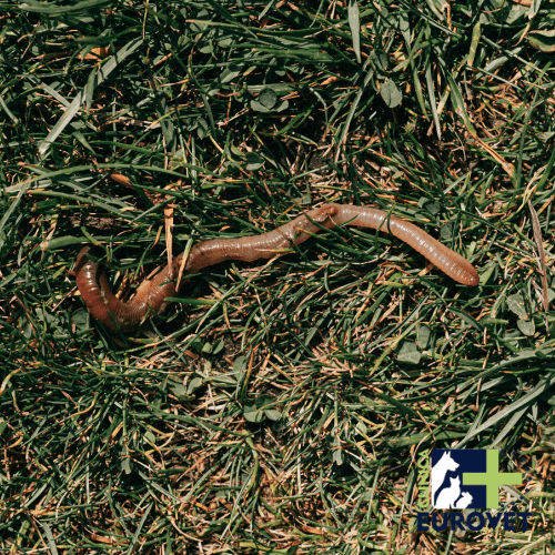
Helminths (worms) in dogs and cats
We classify them mainly into helminths (worms) and protozoa (microscopic parasites). Each group has its own weapons and strategies. Here we present them with their scientific names—nothing is more professional than calling things by their proper names—but explained in simple terms so that you can control the situation like any responsible owner. Supplementing this information with regular coprological examinations (faecal analysis) at least once or twice a year, or more frequently in puppies and high-risk animals, allows you to stay ahead of many problems before they arise, as recommended by the European guidelines for parasite control (ESCCAP).
Helminths are multicellular internal parasites that colonise the digestive tract of dogs and cats and, in many cases, other organs during their life cycle. They are particularly common in puppies and animals that are not regularly dewormed. They are broadly divided into two groups: nematodes, which have a cylindrical body, and cestodes, which have a flat, segmented body. Although many owners only think of “worms in the faeces”, the reality is that these parasites can cause anything from mild digestive disorders to severe anaemia, growth retardation, lung damage or zoonotic problems that affect the family. Their enormous reproductive capacity, the resistance of their eggs in the environment, and the possibility of vertical transmission or transmission through intermediate hosts mean that they must be taken very seriously in modern preventive veterinary medicine. To put a figure on this “environmental resistance,” a global meta-review estimated that, globally, about 1 in 5 public spaces sampled have Toxocara eggs in the soil (pooled prevalence ~21%), which explains why parks and gardens can become persistent sources if transmission chains are not broken.
“BARF” and surprises under the microscope
In a study of 89 animals fed a raw diet (BARF), some “positives” in faeces were not parasites from the animal, but traces from what they ate: Dicrocoelium dendriticum eggs were detected in 6 and Fasciola hepatica DNA in 1. Moral: diet can “contaminate” the diagnosis… and also the environment if hygiene is poor.
Source: https://pmc.ncbi.nlm.nih.gov/articles/PMC11201496/
Nematodes, roundworms that can travel throughout the entire body
Nematodes are cylindrical worms that, in their adult stage, are mainly found in the small intestine, although some species migrate through the liver, lungs or other tissues. They reproduce by means of microscopic eggs that are eliminated in faeces and, once in the environment, can remain infectious for months or even years. This resistance explains why gardens, parks and walking areas easily become parasitic “egg banks” that are difficult to eradicate. Studies and reviews on environmental contamination describe how Toxocara eggs can remain viable in the soil for years, especially in moist and shaded soils, which gives an idea of the “memory effect” of the soil (and why a site that has been “contaminated once” can continue to cause problems long afterwards). For example, a study of public parks found that even when the prevalence of positive parks was relatively low (7.6%), the viability of the eggs detected could be extremely high (≈94%), i.e., “few, but very much alive.”
In puppies, whose immune response is still immature, nematode infestations can quickly destabilise their general condition and compromise their growth. That is why we recommend starting internal deworming at a very early age and repeating it according to the schedule prescribed by the vet, without skipping doses “because the animal seems to be fine”. And, when it comes to diagnosis, technical guidelines remind us that a standard coprology is best interpreted when a sufficient amount of sample is analysed (e.g. 3–5 g of faeces) and when it is repeated if there is clinical suspicion.
From a public health perspective, some of these parasites are zoonotic, meaning that the problem extends beyond the animal to the family environment.
Toxocara spp., the most important intestinal parasite in dogs and one of the most dangerous for humans
Toxocara canis in dogs and Toxocara cati in cats are probably the most important roundworms in small animal practice. Adult females can produce up to 200,000 eggs per day, which are eliminated in the faeces and become infective in the environment after a period of maturation.
These eggs have an extremely resistant shell, capable of protecting the larvae inside for years in soil, sand or grass (especially if there is moisture and shade). When a dog or cat ingests infectious eggs, the larvae emerge in the intestine, pass through the intestinal wall and travel through the body via the liver and lungs, causing inflammation, coughing and, in very young animals, breathing difficulties.
After this migration, the larvae ascend through the trachea, are swallowed and settle in the small intestine, where they complete their development into adult worms that consume nutrients, irritate the mucosa and cause symptoms such as diarrhoea, vomiting, a distended ‘barrel-like’ abdomen and growth retardation due to malabsorption. In adult bitches, some of the larvae may remain “dormant” in tissues and reactivate during pregnancy, crossing the placenta and infecting the foetuses before birth. They can also reach puppies through milk during lactation, which explains why many litters are born with a heavy parasite load.
In humans, especially in children who play on contaminated soil, Toxocara can cause Visceral or Ocular Larva Migrans syndromes, with hepatic, neurological or retinal involvement which, in severe cases, can lead to permanent vision loss. To ground the issue in real data (and not just theory): in Spain, serological studies have been published that show highly variable prevalence rates depending on the area and population; for example, a classic study found seropositivity of 3.6% in adults in Madrid and 17.4% in adults in Tenerife, and in children 0% (Madrid) versus 4.2% (Tenerife). These numbers do not mean “serious illness” in all cases, but they do indicate environmental exposure and justify constant prevention measures.
For all these reasons, it is considered a parasite of utmost importance in public health and a compelling reason to establish strict and consistent deworming programmes. Simple measures such as preventing children from playing in sandboxes where dogs or cats have access, teaching them to wash their hands after playing outdoors, and always picking up faeces in public areas drastically reduce the risk.
Toxascaris leonina, less aggressive than Toxocara, but just as persistent in the environment
Toxascaris leonina is another intestinal nematode that affects both dogs and cats, especially in environments where both species coexist. Unlike Toxocara, its biological cycle is simpler: the larvae do not migrate extensively through the tissues, but develop in the intestine itself after ingestion of infective eggs or paratenic hosts, such as small rodents. This absence of systemic migration usually results in somewhat less severe clinical symptoms, but these are nonetheless significant.
Toxascaris eggs, although somewhat less resistant than those of Toxocara, can also survive for long periods in the environment, especially in damp and shady areas. Their rapid ability to become infectious after being eliminated in faeces facilitates reinfection in homes, shelters and breeding facilities where hygiene is not rigorous. From a zoonotic point of view, Toxascaris is considered to be of much less importance than Toxocara, but its presence in faecal samples is a clear warning sign: it indicates that the animal is chronically exposed to contaminated faeces or infected prey.
Ancylostoma spp., the intestinal vampire that feeds on blood and causes anaemia
Ancylostoma caninum in dogs and Ancylostoma tubaeforme in cats are small nematodes but with a potentially very serious impact. Their mouth capsule is equipped with plates or teeth with which they attach themselves firmly to the mucosa of the small intestine and pierce the capillaries to feed on blood. Each worm can consume several microlitres of blood per day; in heavy infestations, especially in puppies, this sustained loss quickly leads to severe anaemia, with pale mucous membranes, apathy, difficulty exercising and, in extreme cases, collapse. To put this in figures, a recent review summarises losses due to A. caninum in the order of 10–200 µL of blood per worm per day, and the MSD Veterinary Manual cites estimates of up to 0.1 mL per worm per day.
Furthermore, they do not just suck blood: their mechanical action causes microhaemorrhages and inflammatory lesions that promote diarrhoea, sometimes dark in colour due to digested blood.
Ancylostoma larvae can penetrate through the skin, especially through the footpads and areas of thin skin in direct contact with contaminated soil. After penetrating the skin, they can migrate to the lungs and then to the intestine, thus completing a cycle that does not even require the ingestion of eggs. In humans, these larvae are responsible for a condition known as cutaneous larva migrans, characterised by reddish, snaking lines on the skin that cause intense itching.
Uncinaria stenocephala, a parasite adapted to temperate climates and moist soils
Uncinaria stenocephala belongs to the same family as Ancylostoma, but has some differences that are worth noting. Its mouthparts are adapted to attach and feed in the small intestine, although it usually extracts less blood per individual than Ancylostoma, so the anaemia it causes is generally more moderate. To compare with numbers: in the review cited above, U. stenocephala is associated with much lower losses (on the order of 0.3 µL per worm per day, compared to A. caninum).
Even so, in weakened or very young animals, a significant infestation can contribute to weakness, soft stools or chronic diarrhoea and weight loss. From an epidemiological point of view, Uncinaria is more commonly associated with temperate and cool climates, but above all with moist, poorly drained soils, which means that parks, gardens and shaded areas where dogs are regularly present can act as veritable reservoirs.
Trichuris vulpis, the “whipworm” that specialises in the large intestine and causes chronic colitis
Trichuris vulpis, known as the “whipworm” due to its characteristic morphology, is mainly located in the caecum and colon. Its thin, elongated anterior part embeds itself deeply in the intestinal mucosa, while the thicker posterior portion remains free in the intestinal lumen. This form of attachment causes persistent inflammation that results in chronic colitis, with long-term diarrhoea, the presence of mucus and fresh blood in the stool, pain during defecation, and tenesmus, i.e., repeated and painful straining to pass small amounts of faecal matter.
The Trichuris cycle is relatively slow: it can take approximately 74–90 days (≈ 10–12 weeks) from the ingestion of the eggs to the appearance of eggs in the stool, which means that an early coprological test may be falsely negative.
Its eggs, which are barrel-shaped and have characteristic polar caps, have a particularly thick shell that allows them to remain viable for years in the environment. Under optimal conditions, prolonged environmental survival (up to 12 years in soil) has even been reported in technical pest control information material, which explains why a garden can continue to ‘test positive’ season after season if hygiene practices are not changed.
Due to this combination of slow cycle, environmental persistence and chronic digestive signs, Trichuris requires prolonged antiparasitic treatments, repeated coprological checks and, very often, modifications in environmental management to achieve a true resolution of the problem.
Other nematodes of veterinary importance
Although Toxocara, Toxascaris, Ancylostoma, Uncinaria, and Trichuris are the usual protagonists in daily clinical practice, there are other nematodes that can affect dogs and cats and that should be kept in mind. Strongyloides stercoralis, for example, is a parasite with a particularly complex life cycle, with free-living stages in the environment and the ability to produce internal autoinfection, i.e. new generations of parasites without the need for external exposure. In immunocompromised animals, it can trigger severe diarrhoea and systemic symptoms that are difficult to control. Some species of Capillaria can be found in both the intestine and the respiratory tract, leading to chronic respiratory signs that can be confused with bronchitis or other primary lung conditions.
Strongyloides: the “underestimated” parasite in immunosuppression
In addition to the “classics”, in immunocompromised individuals it is important not to underestimate less commonly discussed parasites such as Strongyloides. Recent reviews discuss the role of dogs in zoonotic cycles of Strongyloides stercoralis, which is particularly relevant due to its potential impact on vulnerable individuals (those who are immunocompromised). It is not the typical “park worm”, but it deserves clinical attention.
Source (URL):https://royalsocietypublishing.org/doi/10.1098/rsos.171168
Detection of these “less classic” nematodes requires careful interpretation of coprological tests and often repeat testing or the use of specific techniques.
Cestodes, segmented flatworms that attach themselves to the intestine
Cestodes are flat, segmented helminths consisting of a head called a scolex and a chain of proglottids or segments that grow continuously from the area closest to the scolex towards the tail. The scolex is equipped with suckers and, in many species, hooks that act as a firm anchor in the mucosa of the small intestine. Unlike nematodes, cestodes do not have their own digestive tract: they absorb nutrients directly through their body surface. Their reproductive strategy is based on each proglottid functioning as an almost independent reproductive unit, capable of producing and releasing hundreds or thousands of eggs. In practice, this means that for each adult worm, environmental contamination can be massive. To complete their life cycle, they always need an intermediate host, which can be a flea, a rodent, a rabbit, or even contaminated raw meat. Controlling these intermediate hosts (fleas, rodents, access to carrion) is as important as administering the appropriate antiparasitic treatment.
Taenia spp., parasites transmitted by hunting, carrion and the ingestion of raw meat
The different species of Taenia that affect dogs and cats share a fundamental trait: their life cycle involves the ingestion of larvae encysted in the tissues of intermediate hosts. When a dog hunts a rabbit or a cat consumes a rodent, it is potentially ingesting cysticerci, which are the encapsulated larval forms of Taenia. Once inside the dog or cat’s intestine, these cysticerci evaginate, attach their scolex to the mucosa and begin to produce proglottids. Biological cycle data sheets describe, for example, that Taenia taeniaeformis in cats can reach up to 70 cm and that its prepatent period is 5–6 weeks.
Clinically, tapeworms tend to go largely unnoticed. Many parasitised animals do not show any striking digestive signs, or only have occasional episodes of loose stools, vague abdominal discomfort or reduced exercise tolerance. However, the environmental impact of an adult Taenia is enormous.
Dipylidium caninum, the most common intestinal parasite in pets, linked to fleas
Dipylidium caninum is by far the most common cestode in domestic dogs and cats, and its success is due to a very effective alliance with fleas. When these fleas complete their development and jump onto the animal, they become the key link in the cycle. The dog or cat, scratching or biting due to the itching caused by the bites, ends up ingesting an infected flea.
“Rice grains” with ID
Dipylidium caninum proglottids may look like pumpkin seeds when fresh and rice grains when dry; sometimes they can even be seen “hanging” from the anus. This visual detail is often the quickest diagnostic clue.
Animals parasitised by Dipylidium often show few specific clinical signs. The problem is not usually explosive diarrhoea, but a much more subtle picture of slight digestive discomfort and, above all, anal itching. To relieve this, many dogs perform the typical “scooting” behaviour, dragging their rear end across the floor. Owners may notice small white fragments, resembling grains of rice, moving around the perianal area, on the bed or on the sofa; these are proglottids filled with eggs that, when dry, break open and release their contents into the environment. This description matches the CDC’s DPDx fact sheet, which states that proglottids are “pumpkin seed-like” when fresh and often “resemble grains of rice” when dry; it also describes a typical mature proglottid size of approximately 12 mm × 3 mm.
An important detail for the local context: a review of Spain reports that D. caninum infestations in animals can be frequent and highly variable depending on the study, with published ranges of 8.3–39.0% in dogs and 4.2–64.6% in cats.
In young children who live closely with pets and tend to put their hands in their mouths, cases of Dipylidium infection have been reported after accidentally ingesting a flea. Although the disease is rarely serious in humans, this fact highlights the zoonotic nature of the parasite and the importance of integrated control. Proper treatment does not consist solely of administering an internal antiparasitic to eliminate the worm: it is essential to combine intestinal deworming with a rigorous flea control protocol, both on the animal and in the home environment.
European Scientific Counsel Companion Animal Parasites (ESCCAP). (2025) – Worm Control in Dogs and Cats (ESCCAP Guideline 01, Seventh Edition) – ESCCAP Guidelines – Evidence-based guide for the control of helminths in dogs and cats (Europe). – https://www.esccap.org/uploads/docs/biu0jhej_0778_ESCCAP_GL1__English_2025_v21_1p.pdf
Fakhri, Y., Gasser, R. B., Rostami, A., et al. (2018) – Toxocara eggs in public places worldwide: A systematic review and meta-analysis – Environmental Pollution – Global meta-analysis on the presence of Toxocara eggs in public spaces (soil/parks). – https://pubmed.ncbi.nlm.nih.gov/30142562/
Nava, L. P., Ayerdi, E., Ruiz, J., et al. (2020) – Prevalence and Viability of Toxocara spp. Eggs in Soil of Public Parks in Northwestern Mexico – Iranian Journal of Parasitology – Study of park contamination and viability of Toxocara eggs. – https://pmc.ncbi.nlm.nih.gov/articles/PMC7536377/
Ruiz de Ybáñez, M. R., Garijo, M. M., Alonso, F. D. (2001) – Prevalence and viability of eggs of Toxocara spp. and Toxascaris leonina in public parks in eastern Spain – Parasitology Research – Study in parks in eastern Spain on the prevalence and viability of eggs. – https://link.springer.com/article/10.1007/s004360100409
Public Health Agency of Canada. (2010) – Pathogen Safety Data Sheets: Infectious Substances — Toxocara canis / Toxocara cati – Government of Canada – Official data sheet with information on transmission, risk and environmental survival. – https://www.canada.ca/en/public-health/services/laboratory-biosafety-biosecurity/pathogen-safety-data-sheets-risk-assessment/toxocara-canis.html
Magnaval, J.-F., Glickman, L. T., Dorchies, P., Morassin, B. (2001) – Highlights of Human Toxocariasis – The Korean Journal of Parasitology – Clinical and epidemiological review of human toxocariasis. – https://www.koreamed.org/SearchBasic.php?RID=1016KJP/2001.39.1.1
Liptáková, M., Schreiberová, A., Cellengová, Z., Kožárová, V., Štrkolcová, G. (2025) – The Canine Hookworm Ancylostoma caninum: First Confirmed Evidence in a Dog in Central Europe: Epidemiological Relevance or Coincidence? – Pathogens – Article with quantitative data (e.g., blood loss due to worms) and epidemiological discussion. – https://doi.org/10.3390/pathogens14121241
MSD Veterinary Manual. (2024) – Hookworms in Small Animals – MSD Veterinary Manual – Clinical review (pathogenesis, signs, control) of hookworms in small animals. – https://www.msdvetmanual.com/digestive-system/gastrointestinal-parasites-of-small-animals/hookworms-in-small-animals
Companion Animal Parasite Council (CAPC). (2025) – Trichuris vulpis – CAPC Guidelines – Practical guide (includes prepatent period 74–90 days, environmental viability, etc.). – https://capcvet.org/guidelines/trichuris-vulpis/
ESCCAP UK & Ireland. (n.d.) – Life cycle of the dog whipworm, Trichuris vulpis – ESCCAP UK Parasite Life Cycles – Infographic (includes egg survival up to 12 years in soil under optimal conditions and prepatent period). – https://www.esccapuk.org.uk/link-document/122/
ESCCAP UK & Ireland. (n.d.) – Life cycle of Taenia taeniaformis of cats – ESCCAP UK Parasite Life Cycles – Infographic of the cycle (includes prepatent period of 5–6 weeks). – https://www.esccapuk.org.uk/link-document/121/
Centres for Disease Control and Prevention (CDC). (2019) – DPDx — Dipylidium caninum – CDC DPDx – Official resource (life cycle, diagnosis, measures and morphological data). – https://www.cdc.gov/dpdx/dipylidium/index.html