- Clinica Veterinaria Eurovet - Avenida Estación, 38 - 12598 - Peñiscola
- +34 964 784 983
- +34 671 281 025
- clinica.eurovet@gmail.com



When childbirth becomes complicated
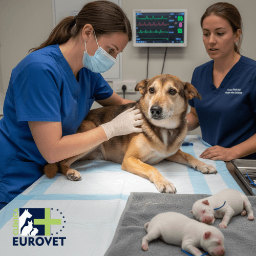
Factors that can turn “natural” into an emergency
Childbirth is a physiological process, yes… but biology has its creative days. In clinical practice, between 5 and 15% of bitches experience some degree of dystocia, a figure that can exceed 20% in certain brachycephalic breeds. In other words, in certain contexts, one in five births may require veterinary assistance. Dystocia is defined as a difficult or impossible birth without intervention, and is one of the most common obstetric emergencies in veterinary medicine.
The good news is that most dystocias do not occur unpredictably. Various retrospective studies agree that a high proportion of cases have identifiable risk factors during pregnancy, and recognising these allows decisions to be made in advance (close monitoring, control of the timing of delivery or planning a caesarean section), reducing maternal complications and improving neonatal survival. For the guardian, the most practical measure is simple: before the last third of gestation, it is advisable to talk to the veterinarian to agree on “what is considered normal” in their case (breed, estimated litter size, history) and to have a contact and transport plan ready. In obstetrics, the difference between “we’ll see” and “emergency” is sometimes a matter of minutes.
In large population studies of insured dogs, dystocia is not an exceptional phenomenon: around 2% of dogs experienced at least one episode of dystocia during their reproductive life and, of those cases, almost two-thirds required a caesarean section. This reinforces an important clinical idea: a significant proportion of complicated births cannot be resolved without intervention and it is advisable to be prepared.
The mother is in charge: maternal factors that can slow down labour
From a physiological point of view, birth depends on two main factors: the uterine motor (effective contractions) and the mechanical motor (a birth canal with sufficient space and no obstacles). When either of these fails, progress stops. Under normal conditions, the expulsion phase should show effective and progressive contractions; when this does not occur, the risk of dystocia increases over time.
At home, the most useful thing is to observe without interfering: a calm environment, without visitors, without children around and with a stable birthing area (large box, clean sheets, comfortable temperature) reduces stress. Stress is not innocent: it can slow down the release of endogenous oxytocin, which is one of the physiological “keys” that sustain the dynamics of labour.
Did you know?🧠
During a normal birth, there may be relatively long pauses between the birth of one puppy and the next, without this implying a problem. The warning sign is not the isolated pause, but the combination of active contractions, lack of progress, and deterioration in the mother’s general condition. It is this triad that warrants immediate evaluation.
Maternal age is a well-documented factor. In older bitches, a decrease in myometrial contractility (the ability of the uterine muscle to contract with force and coordination) has been described, with a higher incidence of primary uterine inertia. In practical terms, late primiparous bitches tend to tolerate long labour less well, and here it is important not to “normalise” the lack of progress: if labour does not progress, the neonatal prognosis worsens the longer intervention is delayed.
In retrospective clinical reference series, analysis of cases of canine dystocia shows that the interval between the onset of the problem and veterinary intervention is one of the most important determinants of neonatal prognosis. In veterinary obstetrics, “waiting a little longer” rarely works in your favour when labour has already stopped progressing.
Among metabolic factors, peripartum hypocalcaemia is particularly important. Calcium is essential for muscle contraction, and low levels are associated with weak or uncoordinated contractions; in practice, this explains why a mother may show effort without being able to expel her offspring. Even so, it is not recommended to supplement calcium “as a matter of course” during pregnancy without veterinary advice, as this can alter physiological regulation. It is useful for the guardian to watch for signs of alarm around the time of birth (tremors, stiffness, heavy panting, disproportionate restlessness) and consult a vet without delay.
Hypocalcaemia in bitches: one piece of the puzzle, not the whole puzzle 🧩
Although hypocalcaemia is often associated with uterine inertia, clinical studies show that not all bitches with inertia have low calcium levels. In other words, calcium may be involved, but dystocia is multifactorial and it is best to avoid “single cause” explanations without a complete evaluation.
From an anatomical point of view, a narrow pelvic canal or bone malformations can prevent delivery even with normal contractions. Obstetric reviews describe foetal-pelvic disproportion as responsible for a significant proportion of dystocia cases, compounded by internal adhesions or scarring (previous surgery, inflammation, trauma) that reduce the distensibility of the birth canal. In females with a history of dystocia or relevant surgery, anticipation is usually the wisest course of action.
Uterine inertia is one of the most common causes of dystocia. It can be primary (the uterus does not initiate effective contractions) or secondary (contractions are exhausted after prolonged effort). Overall, uterine inertia accounts for a significant proportion of canine and feline dystocia, and its prognosis worsens with maternal exhaustion.
Body condition is also a determining factor. Obesity is associated with longer labours, greater fatigue and a higher probability of intervention; at the opposite extreme, malnutrition reduces the energy reserves necessary for effective labour. Added to this are systemic diseases (endocrine, cardiac, inflammatory) that reduce the mother’s tolerance to the physiological stress of labour. In practical terms, in the last two weeks it is advisable to ensure access to fresh water, a digestible and divided diet if she eats little, uninterrupted rest and to avoid “home remedies” to speed up labour: indiscriminate manipulation can increase pain, stress and the risk of infection.
The offspring also play a role: foetal factors that hinder expulsion
From a foetal point of view, the relationship between the size, number and presentation of the offspring is key. Excessive foetal size is a classic cause of dystocia, especially in small litters: with less intrauterine competition for space and nutrients, each puppy can develop more than usual. In other words: small litters, large offspring.
At home, a valuable practical tip is to keep a simple record from the start of the process: the time when restlessness/panting began, the start of active contractions, the birth of each puppy and the general appearance of the mother. This record greatly improves the quality of communication with the vet.
Congenital malformations, although less common, can block the birth canal. They should be suspected when there are effective contractions without progression. For the guardian, here is a golden rule: avoid strong traction or repeated manoeuvres without training. If part of the foetus is visible and it is not progressing with contractions, the safest thing to do is to contact someone, remain calm and avoid persistent manipulation.
Abnormal foetal presentation is another major cause. Longitudinal cephalic presentation with correct alignment is the most favourable; transverse presentations, caudal presentations with poor extension or with flexed limbs can cause blockage and require obstetric intervention or caesarean section.
In cats, dystocia has particular characteristics. In retrospective series, maternal factors predominate over foetal factors, and medical treatment has a limited success rate that depends on the type of dystocia.
Feline dystocia: the mother is often the key 🔍
In cats, retrospective clinical data indicate that dystocia is more often due to maternal factors (such as uterine inertia or birth canal problems) than to foetal problems. This approach is important because it explains why medical management is not always sufficient and why caesarean section continues to play a central role in the management of these patients.
In recent studies, medical treatment with calcium and/or oxytocin has shown acceptable efficacy, especially in non-obstructive dystocia and when few foetuses remain to be born. When there is mechanical obstruction or the process is advanced, the probability of success of medical treatment drops sharply. This idea avoids false expectations and, above all, avoids harmful delays.
Both the presence of a single large foetus and a large litter with poor alignment increase the duration of labour and the risk of maternal exhaustion. It has been clinically observed that when the interval between births is excessively long, especially if there are active contractions without result, the risk of foetal distress increases.
In prolonged births, the greatest risk is not always to the first foetuses, but to the last. As labour progresses, maternal exhaustion and loss of contraction efficiency increase the risk of foetal hypoxia. Therefore, dystocia that is not resolved in time often compromises the last foetuses in the litter in particular.
How to reduce scares: practical (real) prevention of dystocia
Prevention begins during gestation. From approximately day 45 onwards, ultrasound scans allow viability and development to be assessed, while X-rays in the last third facilitate foetal counting and estimation of relative size. Knowing how many offspring are expected reduces the risk of overlooking foetal or placental retention, a classic cause of postpartum complications.
In females with risk factors, pelvic evaluation and analysis of reproductive history help to anticipate mechanical problems. In certain contexts, a planned caesarean section is not giving up: it is preventive medicine. In predisposed breeds, clinical practice shows high rates of caesarean section precisely to avoid emergencies with exhausted mothers or compromised foetuses.
Your vet advises you 📓
Before delivery, it is advisable for the guardian to be clear about three things: approximately how many puppies are expected, what time thresholds or signs are considered abnormal in their case, and who to call if questions arise. In obstetrics, consulting early is never a mistake, but consulting late can be.
Finally, active veterinary monitoring during birth has a direct impact on the prognosis. Early intervention reduces perinatal mortality and maternal complications; waiting too long when there are clear signs of dystocia worsens the outcome. A practical detail that helps a lot is to communicate observable data rather than general impressions: “has been in active labour for X amount of time,” “last birth was X,” “there is effort without expulsion,” “she is exhausted,” “there is abnormal discharge.” This allows for a more accurate assessment of the urgency. 📞
Key idea
For years, childbirth was considered a “natural” process that should be interfered with as little as possible. Modern veterinary obstetrics has nuanced this view: natural processes can also become complicated, and prevention based on assessment, monitoring, and planning is now considered high-quality medicine, not excessive interventionism.
Between 5 and 15% of births can be complicated (and more in certain cases), but a significant proportion of dystocias have identifiable risk factors. With information, monitoring and planning, many emergencies can be avoided or resolved in time. Dystocia rarely occurs without warning. Ultrasound, X-rays, analysis of medical history and a clear plan of action make the difference between a controlled birth and a life-threatening emergency. Waiting for “something to go wrong” usually means arriving too late.
Bergström, A., Nødtvedt, A., Lagerstedt, A. S., Egenvall, A. (2006) – Risk factors and frequency of dystocia and caesarean section in bitches: a population-based study of insured animals in Sweden. – Journal of Small Animal Practice – Large-scale epidemiological study analysing the incidence of dystocia, caesarean section rates and breed predisposition in bitches. – https://pubmed.ncbi.nlm.nih.gov/17187641/
Darvelid, A. W., Linde-Forsberg, C. (1994) – Dystocia in bitches: retrospective study of 182 clinical cases. – Journal of Small Animal Practice – Classic clinical analysis describing the maternal and foetal causes of dystocia and emphasising the importance of early diagnosis and intervention. – https://onlinelibrary.wiley.com/doi/abs/10.1111/j.1748-5827.1994.tb03863.x
Bailin, S., Axnér, E., Gustavsson, H., Linde-Forsberg, C. (2022) – Dystocia in cats: causes, treatment and clinical outcome in a retrospective study. – Journal of Feline Medicine and Surgery – Study showing the predominance of maternal factors in feline dystocia and the limited effectiveness of medical treatment compared to surgery. – https://pubmed.ncbi.nlm.nih.gov/34124965/
Axnér, E., Gustavsson, H., Linde-Forsberg, C. (2025) – Results of medical and surgical management of feline dystocia in 111 retrospective cases. – Journal of Feline Medicine and Surgery – Recent clinical review analysing uterine inertia in cats and defining the conditions under which treatment with calcium and oxytocin may be effective. – https://pubmed.ncbi.nlm.nih.gov/40221772/
Hollinshead, F. K., Hanlon, D. W. (2018) – Serum calcium concentrations and their relationship to uterine inertia in bitches. – Reproduction in Domestic Animals – Study evaluating the association between hypocalcaemia and primary and secondary uterine inertia in bitches with dystocia. – https://onlinelibrary.wiley.com/doi/10.1111/rda.13157