- Clinica Veterinaria Eurovet - Avenida Estación, 38 - 12598 - Peñiscola
- +34 964 784 983
- +34 671 281 025
- clinica.eurovet@gmail.com



Hip dysplasia: clinical signs you should not ignore
When gait says more than words
In senior dogs, hip dysplasia can appear subtly: no cries of pain, but clear signals if you know how to read them. An unusual gait, difficulty getting up, or avoiding play may be the first hints that something is wrong with the joints.
In young dogs (4–12 months), signs are usually intermittent: lameness after exercise, trouble rising, or a “hopping” gait with both hind limbs. In adults, chronic pain causes muscle atrophy, reluctance to move, and behavioral changes.
In cats, hip dysplasia is even more inconspicuous. Some may show mild lameness, but more often stiffness when rising and reduced activity are observed.
Signs not to overlook
There are certain clinical signs that – even if they seem subtle at first – may indicate a serious joint problem and should not be ignored:
• Intermittent lameness, especially after activity or long rest.
• Characteristic “hip sway” while walking – a catwalk style, but without the glamour.
• Arched posture or hind limbs kept close together as a pain compensation.
• Difficulty climbing stairs or jumping into a car, especially in large or older dogs.
• Loss of muscle mass in the hindquarters due to reduced activity.
• Behavioral changes: irritability, avoidance of contact, withdrawal.
•
These signs are often mistaken for the belief that the animal is “just getting old.” In many cases, however, they are symptoms of chronic pain. Do not normalize them! A veterinary checkup can be decisive for quality of life.
👉 If you see one of these signs: don’t wait! Schedule a full orthopedic exam.
Orthopedic exam: how we assess the joint
During consultation, we perform a complete orthopedic palpation to assess pain, range of motion, and stability.
• In puppies, the Ortolani test allows us, through pressure and rotation maneuvers, to provoke subluxation and detect joint laxity.
• In adult animals, stiffness, resistance to extension, or loss of function can be observed.
• In cats, the examination is more subtle, noting indirect signs like reduced jumping ability or pain on forced extension.
Diagnosis: radiographs and functional assessment
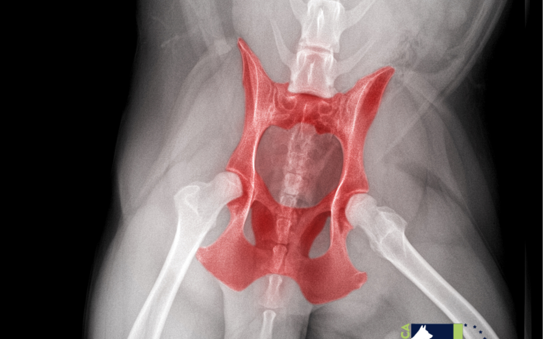
The most reliable method to confirm dysplasia is radiography under sedation, which allows proper positioning and detailed imaging without muscular interference. Evaluated aspects include:
• Joint congruence: how well the femoral head fits into the acetabulum.
• Acetabular coverage: how much of the femoral head is covered by the socket.
• Joint laxity: tendency toward subluxation.
• Degenerative changes: osteophytes, subchondral sclerosis, acetabular remodeling.
The standard reference is the ventrodorsal radiograph with extended limbs.
In some cases, CT or MRI complement the diagnosis.
Functional assessment (gait, daily movements) provides essential information to correlate imaging with clinical signs.
Classification and severity: understanding the situation
Dysplasia is classified by the degree of subluxation and bone changes. This helps with prognosis and treatment planning: from physiotherapy, weight control, and chondroprotectants, to surgeries such as triple pelvic osteotomy or total hip replacement.
FCI classification (Europe, grades A–E):
- Grade A (normal): perfect congruence, round femoral head, deep acetabulum, no dysplasia.
- Grade B (almost normal): slight incongruence, reduced coverage, not yet pathological but monitoring recommended.
- Grade C (mild): clear incongruence, reduced coverage, early remodeling or sclerosis.
- Grade D (moderate): partial subluxation, shallow acetabulum, visible osteophytes.
- Grade E (severe): luxation or severe subluxation, deformed femoral head, shallow acetabulum, advanced osteoarthritis.
PennHIP method:
Especially useful in puppies as young as 16 weeks.
- Values of 0.2–0.3 = good stability.
- Values > 0.7 = high risk of osteoarthritis.
Early diagnosis: a window of opportunity
Early detection, especially in predisposed breeds, changes the entire course of the disease.
- Grades A and B: prevention, risk control, balanced nutrition, moderate exercise, regular checkups.
- Grade C: physiotherapy, strict weight control, possibly preventive surgery in young animals.
- Grades D and E: often surgical solutions (triple pelvic osteotomy in young dogs, total hip replacement in advanced osteoarthritis). If surgery is not possible: intensive pain and inflammation management.
At Eurovet, we recommend preventive checkups at 4–6 months in large breeds, combined with early radiographs. Early detection means better prognosis.